
Why Are Ear Infections Difficult to Diagnose?
Understanding the Limits of the Visual Examination and the Need for More Objective Information
Andrea R. Gravatt, MD, FAAP, FAWM, Professor of Pediatric Medicine, University of Washington School of Medicine (retired status), Clinical Professor of Pediatric Medicine, Elson S. Floyd College of Medicine, Washington State University (retired status)
Key Takeaways
• The middle ear space is located behind the tympanic membrane (eardrum) and cannot be examined directly during conventional visual otoscopy (visual examination with an otoscope).
• Symptoms of acute otitis media (AOM) can be nonspecific, particularly in infants and young children.
• Cerumen, narrow ear canals, patient movement, fluid, and incomplete visualization of the eardrum can interfere with examination and diagnosis.
• Otoscopic interpretation depends on clinician training, technique, and experience, creating potential variability among observers and contributing to incorrect diagnoses and the potential underuse or overuse of antibiotic treatment.
• No routine bedside test currently determines whether AOM is caused by a viral or bacterial infection.
• More objective and reproducible diagnostic technologies may help reduce uncertainty, support appropriate treatment decisions, and prevent complications associated with AOM.
Diagnosing More Than an Earache
Ear infections are among the most common illnesses treated in the pediatric population; however, diagnosing them accurately is not always straightforward. Ear pain, fever, irritability, sleep disruption, and ear pulling can be associated with acute otitis media (AOM), but these symptoms are not specific to a middle ear infection. Some children with AOM have few obvious symptoms, while similar complaints can occur with other illnesses.
The clinician must combine the child’s symptoms and medical history with a physical examination, including a careful evaluation of the ear and the tympanic membrane (eardrum). The clinician also evaluates for middle ear effusion (OME), or fluid behind the eardrum. Each of these steps introduces potential uncertainty, especially when evaluating infants and young children.
The Middle Ear Space
Conventional visual otoscopy (ear examination with an otoscope) allows a clinician to inspect the ear canal and the outer surface of the tympanic membrane. It does not provide a direct view of the middle ear space behind it. The diagnosis of ear pathology therefore requires accurate evaluation of the visible ear structures and assessment of the middle ear space.
Clinicians assess potential ear pathology using information obtained from the medical history, patient symptoms, and physical examination. The physical examination includes attention to:
• The external ear, including the area behind the ear and the scalp
• The appearance of the external ear and ear canal
• The presence of fluid, blood, pus, cerumen, or a foreign body in the ear canal
• The position and contour of the tympanic membrane
• Perforation, bulging, or retraction of the tympanic membrane
• The color and translucency of the tympanic membrane
• Visible fluid, air-fluid levels, or a mass behind the tympanic membrane
• Tympanic membrane mobility
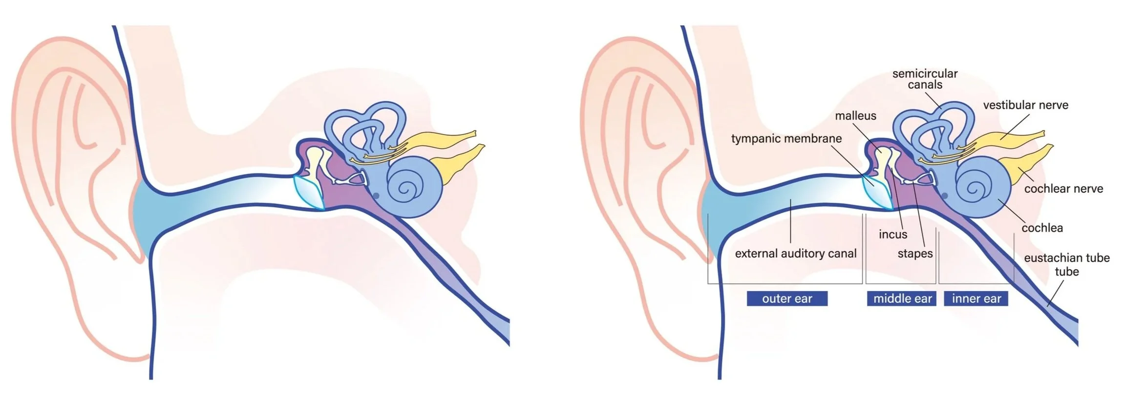
The anatomy diagram below identifies the components of the outer ear, middle ear, and inner ear.
Many of these findings can be subtle. Redness alone, for example, may occur when a child is crying, has a fever, or has irritation unrelated to acute otitis media. Fluid may persist after an infection resolves, and OME can occur without the signs and symptoms of acute infection.
Distinguishing a normal ear from AOM and OME is important because treatment differs among these conditions, and unnecessary antibiotic use should be avoided.

Examining Young Children Can Be Technically Difficult
A comprehensive ear examination may be challenging when the patient is an infant or young child. At times, a child’s developmental stage, sensory processing differences, or severe pain can make it difficult to evaluate the tympanic membrane. Crying, resistance, sudden movement, small ear canals, and difficulty maintaining a stable examination position may interfere with adequate visualization of the tympanic membrane.
Cerumen (earwax) may partially or completely obstruct the ear canal. Narrow or curved external auditory canals may limit the viewing angle. Clinicians may also have difficulty seeing the entire tympanic membrane, making it more difficult to evaluate bulging, fluid, and inflammation.
Accurate Otoscopy Depends on Training and Interpretation
Otoscopy is not simply a matter of looking into the ear. Accurate examination requires appropriate equipment, positioning, visualization, knowledge of ear anatomy, and correct interpretation of physical findings.
Diagnostic performance can vary with training and experience. Published literature reports diagnostic accuracy estimates ranging from 30% to 67.5% for specific pediatric ear diagnoses when primary care assessments were compared with otolaryngologist evaluations.
Differences can also occur among clinicians interpreting the same or similar findings. This interobserver variability may contribute to overdiagnosis, underdiagnosis, and inconsistent treatment decisions.
Middle Ear Fluid Does Not Always Indicate an Acute Infection
Middle ear effusion is fluid located behind the tympanic membrane. The presence or absence of effusion is an important part of evaluating suspected AOM, but fluid alone does not establish the cause or the need for antibiotic therapy.
Fluid may be present:
• During acute otitis media
• After an infection has resolved
• With eustachian tube dysfunction
• Without symptoms of acute infection
A diagnosis of AOM requires more than identifying fluid. Clinicians must interpret the presence of fluid together with the appearance of the tympanic membrane, the onset and severity of symptoms, and established diagnostic criteria.
This distinction is particularly important because OME generally does not require antibiotic treatment, while AOM may benefit from antibiotics.
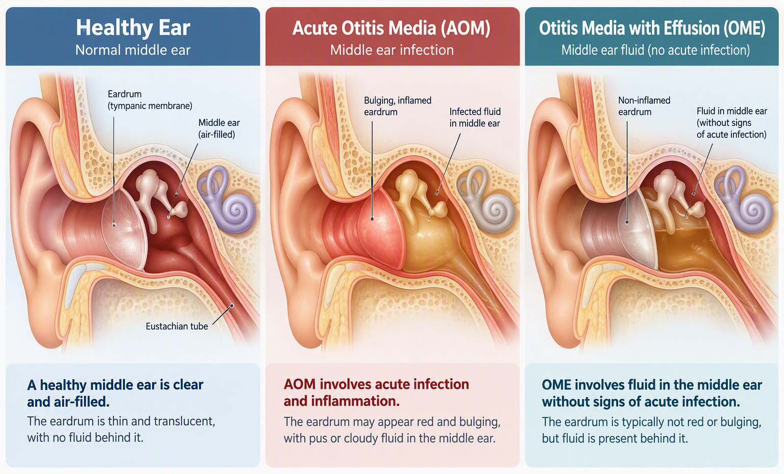
The diagram below illustrates a healthy ear with air behind the eardrum, acute otitis media with infection and inflammation, and otitis media with effusion, which involves fluid in the middle ear without signs of acute infection.
Pneumatic Otoscopy Can Be Valuable, but It Requires Technique
Clinical guidelines recommend pneumatic otoscopy as an important method for assessing suspected middle ear effusion. Unlike standard otoscopy, pneumatic otoscopy introduces gentle changes in air pressure into the ear canal so that the clinician can evaluate eardrum mobility.
Under optimal conditions and in experienced hands, pneumatic otoscopy can perform well in detecting middle ear effusion. However, effective use requires:
• An unobstructed view of the tympanic membrane
• An airtight seal in the ear canal
• Appropriate pressure application
• A cooperative or adequately stabilized patient
• Correct interpretation of eardrum movement
These conditions can be difficult to achieve consistently in busy primary care, urgent care, and emergency settings. Patient movement, an incomplete seal, or subtle changes in tympanic membrane mobility can reduce confidence in the result. Pressure changes may also cause discomfort for some children.
Tympanometry Provides Objective Information, but Not the Whole Answer
Tympanometry evaluates aspects of middle ear function by measuring how the tympanic membrane responds to changes in air pressure. It can provide objective information that supports the detection of middle ear effusion.
Tympanometry does not independently establish a diagnosis of acute bacterial otitis media. The result must be interpreted in conjunction with patient symptoms, otoscopic findings, and the complete clinical presentation.
This illustrates a broader challenge: a tool may help confirm that fluid or abnormal middle ear mechanics are present without identifying whether the underlying condition is acute, resolving, viral, bacterial, or noninfectious.
Viral and Bacterial Infections Can Look Similar
Acute otitis media frequently develops after a viral upper respiratory infection. Viral inflammation can impair eustachian tube function, promote middle ear fluid accumulation, and create conditions that allow bacterial pathogens to enter or proliferate within the middle ear.
Viral and bacterial organisms may coexist. The child’s symptoms and the visual appearance of the tympanic membrane generally reflect the inflammatory response rather than identifying a specific pathogen.
No reliable routine bedside test currently distinguishes viral from bacterial acute otitis media without an invasive procedure. Determining the causative organism requires collection of middle ear fluid. In uncomplicated cases of otitis media, obtaining a sample of middle ear fluid is not routine.
This limitation can make treatment selection difficult. Clinicians need to balance the risk of withholding treatment from a child with progressive bacterial disease against the risks of prescribing antibiotics when they are unlikely to provide benefit.

Diagnostic Uncertainty Affects Antibiotic Decisions
Acute otitis media remains a leading reason for antibiotic prescribing. When the diagnosis is uncertain, clinicians may prescribe antibiotics as a precaution, even when acute bacterial infection is not firmly established.
Antibiotics are important for appropriately selected patients, but unnecessary exposure can contribute to diarrhea, rash, vomiting, allergic reactions, antimicrobial resistance, additional healthcare utilization, and disruption of the developing gut microbiome.
Improved diagnostic confidence could help clinicians better identify children most likely to benefit from treatment while supporting observation when clinically appropriate.
Why Watchful Waiting Also Requires Diagnostic Confidence
Recent clinical pediatric guidelines permit initial observation, often called “watchful waiting”, for selected children with non-severe acute otitis media when reliable follow-up can be ensured.
This approach can reduce unnecessary antibiotic exposure, but it depends upon:
• Confidence in the initial diagnosis
• Appropriate patient selection
• Caregiver understanding
• Access to follow-up
• Timely treatment if symptoms persist or worsen
When the examination is inconclusive, or follow-up is uncertain, implementing watchful waiting may become more difficult. Diagnostic accuracy therefore supports both appropriate antibiotic treatment and the safe use of observation strategies.
The OtoNexus Approach
OtoNexus is developing an investigational ultrasound otoscope that combines proprietary through-air ultrasound sensing with AI-assisted signal analysis.
The system is being designed to collect ultrasound signals through the air-filled ear canal during a routine ear examination. Reflected signals may provide information about the mechanical and acoustic properties of the eardrum and middle ear. Ongoing research is focused on mapping quantitative signal patterns to objective information about middle ear fluid conditions and supporting clinical assessment.
The OtoNexus ultrasound otoscope has not been cleared by the FDA and is not currently available for commercial sale. Final device functionality, workflow, performance characteristics, and indications for use will depend on ongoing development, clinical evaluation, and applicable FDA review.
Looking Ahead
Ear infection diagnosis will continue to require clinical judgment, patient history, physical examination, and evidence-based diagnostic criteria.
The availability of objective, reproducible information about the middle ear could address an important gap in the current examination. Continued advances in sensing, imaging, signal analysis, and artificial intelligence may help clinicians assess middle ear conditions with greater confidence, promote antibiotic stewardship, and improve care for children with suspected ear infections.
Frequently Asked Questions
-
The middle ear is located behind the eardrum. Conventional otoscopy provides a view of the ear canal and tympanic membrane, so clinicians must infer middle ear conditions from eardrum appearance, mobility, symptoms, and other clinical findings.
-
No. Eardrum redness can occur for several reasons and should not be used alone to diagnose AOM. Clinicians evaluate multiple findings, including bulging, mobility, middle ear effusion, symptoms, and medical history.
-
No. Middle ear effusion may occur during an acute infection, remain after an infection resolves, or occur without signs or symptoms of acute infection.
-
Visual otoscopy alone cannot reliably identify the causative organism. Viral and bacterial infections can produce overlapping symptoms and inflammatory findings.
-
Pneumatic otoscopy uses controlled changes in ear-canal pressure to assess eardrum mobility. Reduced mobility may help indicate the presence of middle ear effusion.
-
Accurate diagnosis helps guide appropriate treatment, supports observation when suitable, reduces unnecessary antibiotic exposure, and helps identify children who may require timely intervention.
Medical Disclaimer
This article is provided for educational purposes only and is not intended to replace professional medical advice, diagnosis, or treatment. Patients and caregivers should consult a qualified healthcare professional regarding symptoms or treatment decisions.
References
Shaikh N. Otitis media in young children. N Engl J Med. 2025;392(14):1418-1426. doi:10.1056/NEJMcp2400531.
Folino F, Caruso M, Bosi P, Aldè M, Torretta S, Marchisio P. Acute otitis media diagnosis in childhood: still a problem in 2023? Ital J Pediatr. 2024;50(1):19.
Rosenfeld RM, Shin JJ, Schwartz SR, et al. Clinical practice guideline: otitis media with effusion update. Otolaryngol Head Neck Surg. 2016;154(1 Suppl):S1-S41. doi:10.1177/0194599815623467.
Buchanan CM, Pothier DD. Recognition of paediatric otopathology by general practitioners. Int J Pediatr Otorhinolaryngol. 2008;72(5):669-673. doi:10.1016/j.ijporl.2008.01.030.
Rosenfeld RM. Diagnostic certainty for acute otitis media. Int J Pediatr Otorhinolaryngol. 2002;64(2):89-95.
Lieberthal AS, Carroll AE, Chonmaitree T, et al. The diagnosis and management of acute otitis media. Pediatrics. 2013;131(3):e964-e999. doi:10.1542/peds.2012-3488.
Marom T, Nokso-Koivisto J, Chonmaitree T. Viral-bacterial interactions in acute otitis media. Curr Allergy Asthma Rep. 2012;12(6):551-558.
Sawada S, Okutani F, Kobayashi T. Comprehensive detection of respiratory bacterial and viral pathogens in the middle ear fluid and nasopharynx of pediatric patients with acute otitis media. Pediatr Infect Dis J. 2019;38(12):1199-1203.
Butler AM, Brown DS, Durkin MJ, et al. Association of inappropriate outpatient pediatric antibiotic prescriptions with adverse drug events and health care expenditures. JAMA Netw Open. 2022;5(5):e2214153. doi:10.1001/jamanetworkopen.2022.14153.
Hum SW, Shaikh KJ, Musa SS, Shaikh N. Adverse events of antibiotics used to treat acute otitis media in children: a systematic meta-analysis. J Pediatr. 2019;215:139-143.e7. doi:10.1016/j.jpeds.2019.08.043.
Gerber JS, Jackson MA, Tamma PD, et al. Antibiotic stewardship in pediatrics. Pediatrics. 2021;147(1):e2020040295. doi:10.1542/peds.2020-040295.
McDonnell L, Gilkes A, Ashworth M, et al. Association between antibiotics and gut microbiome dysbiosis in children: systematic review and meta-analysis. Gut Microbes. 2021;13(1):1870402.
Marom T, Kraus O, Habashi N, Tamir SO. Emerging technologies for the diagnosis of otitis media. Otolaryngol Head Neck Surg. 2019;160(3):447-456. doi:10.1177/0194599818809337.
Habib AR, Kajbafzadeh M, Hasan Z, et al. Artificial intelligence to classify ear disease from otoscopy: a systematic review and meta-analysis. Clin Otolaryngol. 2022;47(3):401-413. doi:10.1111/coa.13925.
Red Book: 2024–2027 Report of the Committee on Infectious Diseases (33rd Edition) https://doi.org/10.1542/9781610027373-S4_003_006