
What Is Middle Ear Effusion?
Why fluid develops behind the eardrum, what it means, and how it is diagnosed and managed
Andrea R. Gravatt, MD, FAAP, FAWM, Professor of Pediatric Medicine, University of Washington School of Medicine (retired status), Clinical Professor of Pediatric Medicine, Elson S. Floyd College of Medicine, Washington State University (retired status)
Key Takeaways
Middle ear effusion (MEE) means fluid is present behind the eardrum. It can occur with an acute ear infection (AOM) or remain after the infection has resolved (OME).
Middle ear fluid does not always indicate an active infection. AOM involves fluid with acute inflammation or infection, while OME may involve fluid without signs or symptoms of acute infection.
Children are particularly susceptible to ear infections because their eustachian tubes are still developing. In young children, these tubes are shorter, wider, and more horizontal, which can contribute to problems with middle ear ventilation and drainage. Bacteria and virus particles from the nose and back of the throat can reach the middle ear through the eustachian tube.
Symptoms of OME may be subtle. Some children have few obvious symptoms, but persistent fluid may cause hearing difficulties and subtle changes in listening, language, or school performance.
Accurate diagnosis of OME depends on middle ear evaluation, not symptoms alone. Otoscopy, pneumatic otoscopy and tympanometry can help identify middle ear fluid, while hearing testing can assess its effect on hearing.
Management depends on the type of otitis media, how long the fluid persists, and whether it affects hearing. Many cases of OME resolve, while persistent OME with hearing difficulties may require additional management, including consideration of ventilation tubes (a surgical procedure).
Middle ear fluid occurs in several forms of otitis media, but the cause, symptoms, and appropriate management can differ widely.
Why does fluid develop behind the eardrum?
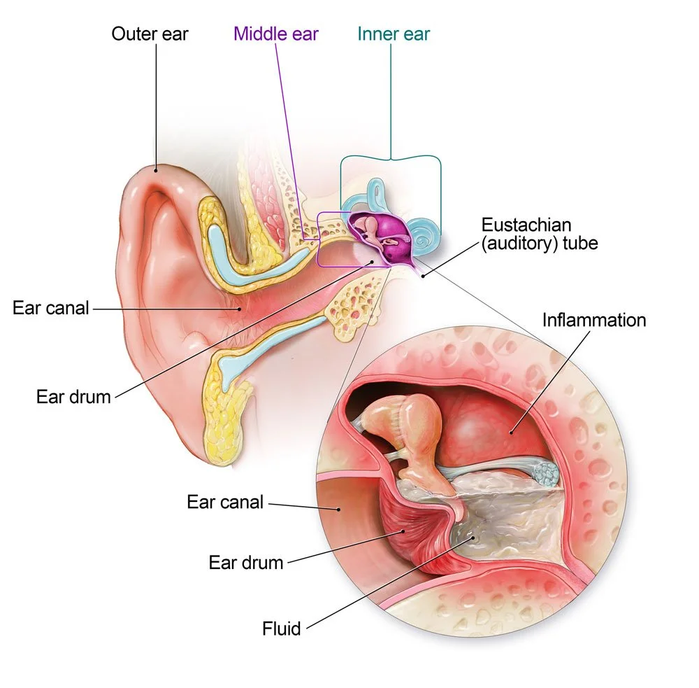
The eustachian tube connects the middle ear space to the back of the nose and throat. It has three important functions: ventilating the middle ear space, helping equalize air pressure, and allowing normal secretions to drain.
Children are especially prone to middle ear problems because their eustachian tubes are shorter, wider, and more horizontal than adults’. These anatomical differences can reduce drainage effectiveness and allow viruses and bacteria from the nose and throat to reach the middle ear more easily. As children grow, the eustachian tube becomes longer and more vertical and generally functions more effectively.
Colds and other upper respiratory infections can inflame the eustachian tube, creating negative pressure and allowing fluid to accumulate. Inflammation can also increase mucus production. Some middle ear fluid becomes thick and sticky, making it more difficult for the ear's normal clearance system to drain.
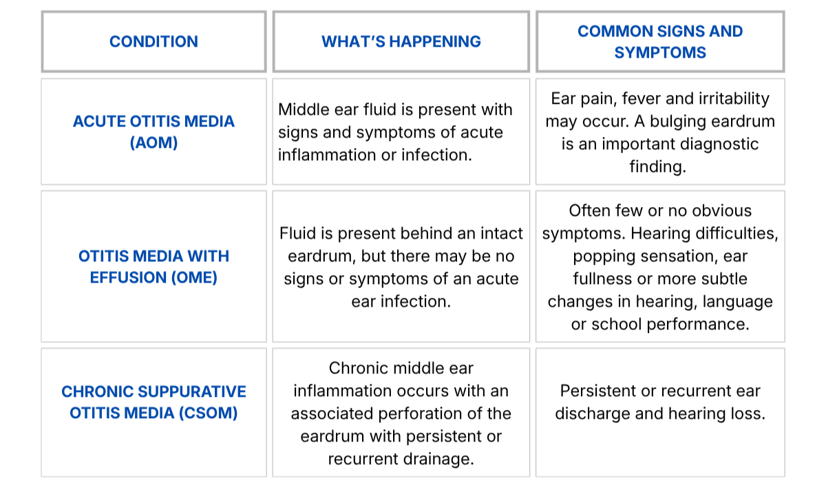
AOM, OME and CSOM: What is the difference?
Otitis media describes several related middle ear conditions. The presence of fluid is important, but fluid alone does not tell us which condition a child has.
Who is more likely to develop otitis media?
Otitis media does not have a single cause. Risk is influenced by a combination of age, anatomy, genetics, recent infections, individual susceptibility and a child's environment.
Factors associated with increased risk include young age, frequent upper respiratory infections, family history of otitis media, craniofacial conditions such as cleft palate, adenoid enlargement, daycare attendance, other siblings, infant bottle propping during feedings, and exposure to tobacco smoke.
Allergic rhinitis has also been associated with OME. Both inflammation and eustachian tube dysfunction are believed to be two factors contributing to the increased incidence seen in these children.
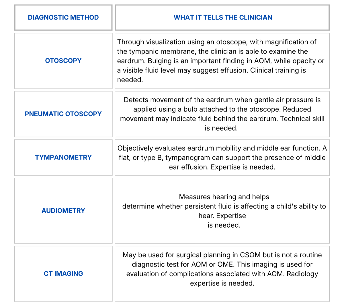
How do clinicians detect middle ear fluid?
Symptoms alone cannot reliably determine whether fluid is present. Examining the eardrum and assessing middle ear function are important parts of diagnosis.
Should children be screened for OME?
Routine screening for OME is not recommended for otherwise healthy children without symptoms, as many episodes resolve naturally. Children at increased risk for speech, language, or learning problems may need closer evaluation and monitoring. Parents’ or caregivers' concerns about a child's hearing should also be taken seriously and evaluated objectively.
How is otitis media managed?
Treatment depends on the type of otitis media, the child's age and symptoms, how long the fluid has been present, hearing status, and other individual risk factors.
AOM: Pain management is an important part of treatment. Depending on age, severity, and other clinical findings, a clinician may recommend observation with close follow-up or antibiotics.
Recurrent AOM: Recurrent AOM is generally defined as three episodes within six months or four within 12 months, with at least one episode during the preceding six months. Current guidelines recommend against tympanostomy tubes when recurrent AOM is present without middle ear effusion at the time of tube placement assessment. When recurrent AOM is accompanied by effusion, bilateral tubes may be offered as a management option.
OME: Many episodes of OME resolve spontaneously. Observation is appropriate for most otherwise healthy children. If OME persists for three months or longer, evaluate hearing. Children with bilateral OME lasting at least three months and documented hearing difficulties should be offered tympanostomy tubes.
What are ventilation tubes?
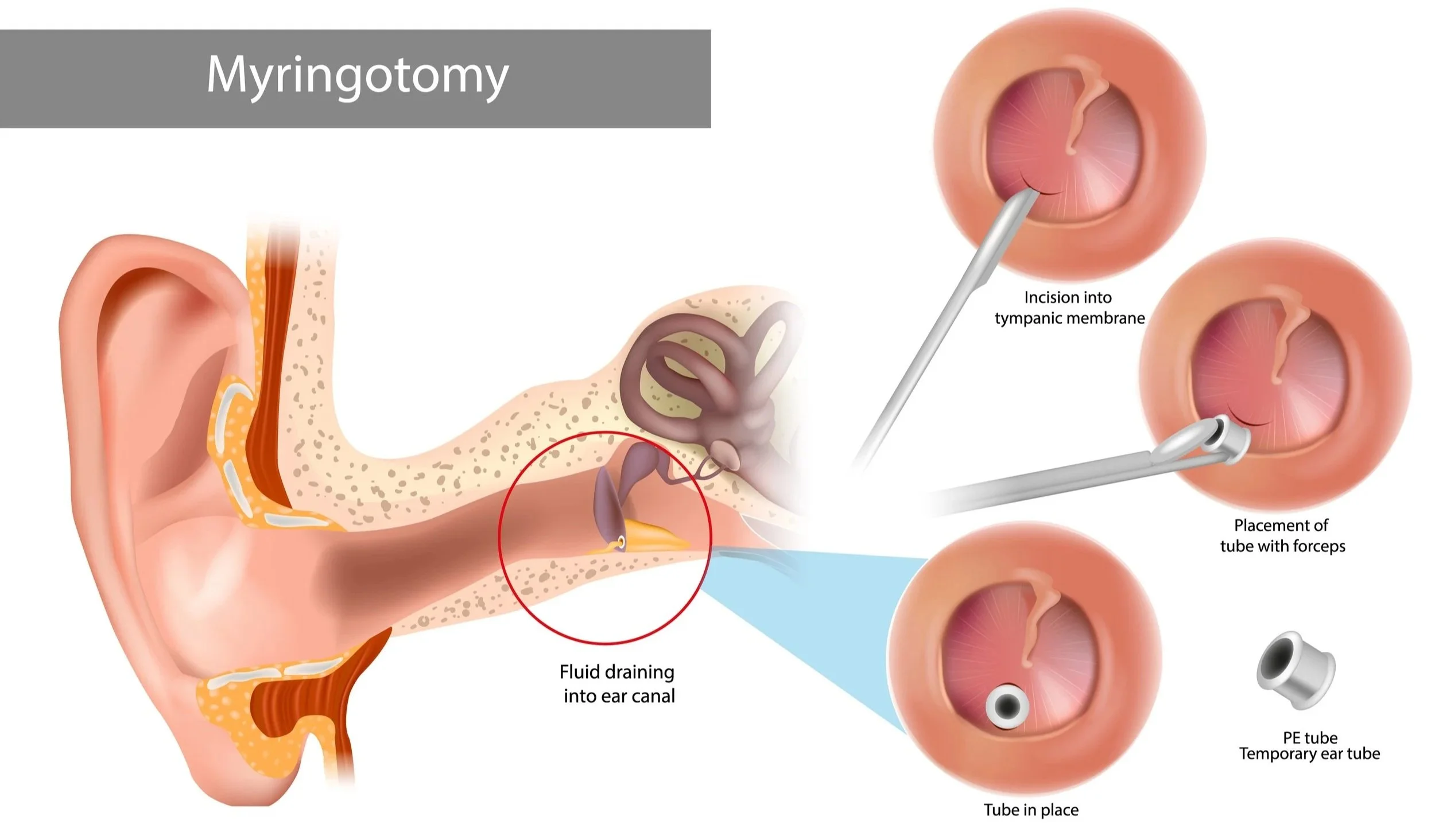
Ventilation tubes, also called tympanostomy tubes or ear tubes (PE tubes), are tiny tubes placed through the eardrum during a surgical procedure (myringotomy). The opening lets air enter the middle ear directly, helping ventilate the space.
This article is provided for educational purposes and is not intended to replace medical evaluation, diagnosis, or treatment by a qualified healthcare professional.
References
Schilder AGM, Chonmaitree T, Cripps AW, Rosenfeld RM, Casselbrant ML, Haggard MP, Venekamp RP. Otitis media. Nat Rev Dis Primers. 2016;2:16063. doi:10.1038/nrdp.2016.63.
Vanneste P, Page C. Otitis media with effusion in children: Pathophysiology, diagnosis, and treatment. A review. J Otol. 2019;14:33-39. doi:10.1016/j.joto.2019.01.005.
Lieberthal AS, Carroll AE, Chonmaitree T, et al. The diagnosis and management of acute otitis media. Pediatrics. 2013;131(3):e964-e999. doi:10.1542/peds.2012-3488.
Wang F, Yu C, Liu R. Causal relationship between allergic rhinitis and otitis media: A Mendelian randomization study. Medicine (Baltimore). 2024 Sep 27;103(39):e39671. doi: 10.1097/MD.0000000000039671. PMID: 39331880; PMCID: PMC11441937.
Dodson KM, Cohen RS, Rubin BK. Middle ear fluid characteristics in pediatric otitis media with effusion. Int J Pediatr Otorhinolaryngol. 2012;76:1806-1809. doi:10.1016/j.ijporl.2012.09.005.
Rosenfeld RM, Tunkel DE, Schwartz SR, et al. Clinical practice guideline: Tympanostomy tubes in children (update). Otolaryngol Head Neck Surg. 2022;166(1_suppl):S1-S55. doi:10.1177/01945998211065662.
Wojas O, Krzych-Fałta E, Furmańczyk K, et al. Co-occurrence of otitis media with effusion and another environment-dependent disease (selected allergic conditions). Adv Dermatol Allergol. 2024;41(1):78-84. doi:10.5114/ada.2023.135602.