
Complications of Otitis Media
Why Recognition and Follow-Up Matter
Andrea R. Gravatt, MD, FAAP, FAWM
Professor of Pediatric Medicine, University of Washington School of Medicine, Clinical Professor of Pediatric Medicine, Elson S. Floyd College of Medicine, Washington State University
Key Takeaways
Serious complications are uncommon in acute otitis media (AOM).
Complications of otitis media may be extracranial or intracranial.
Reported extracranial complications include mastoiditis, subperiosteal abscess, labyrinthitis, and facial nerve involvement.
Reported intracranial complications include meningitis, brain abscess, venous sinus thrombosis, and empyema.
Middle-ear effusion can persist after AOM symptoms resolve, and current guidelines recommend hearing evaluation when OME persists for at least three months, or at any duration in an at-risk child.
Otitis media (OM) is a common childhood infection, and most episodes resolve without serious consequences. These infections are caused by viruses, bacteria, or both. In some cases, however, middle-ear infections may be associated with persistent effusion (fluid) and associated hearing loss. Speech and developmental delays may develop if recurrent ear infections occur in early childhood. In more severe cases of OM, infection may extend beyond the middle-ear space to cause infections in the inner ear space, intracranial and extracranial structures. These more serious complications of OM, including sinus infections, brain abscess, mastoiditis, and bone infections, require hospitalization and possible surgical intervention.
Although vaccines, antibiotics, improved imaging, and multidisciplinary management have changed the clinical landscape, complications of acute and chronic otitis media continue to be reported. Understanding these complications underscores the importance of accurate diagnosis, appropriate management, and clinical follow-up.
How Can Otitis Media Lead to Complications?
The literature describes complications of otitis media based on whether infection extends outside or within the cranial cavity, classifying them as extracranial, intracranial, or combined.
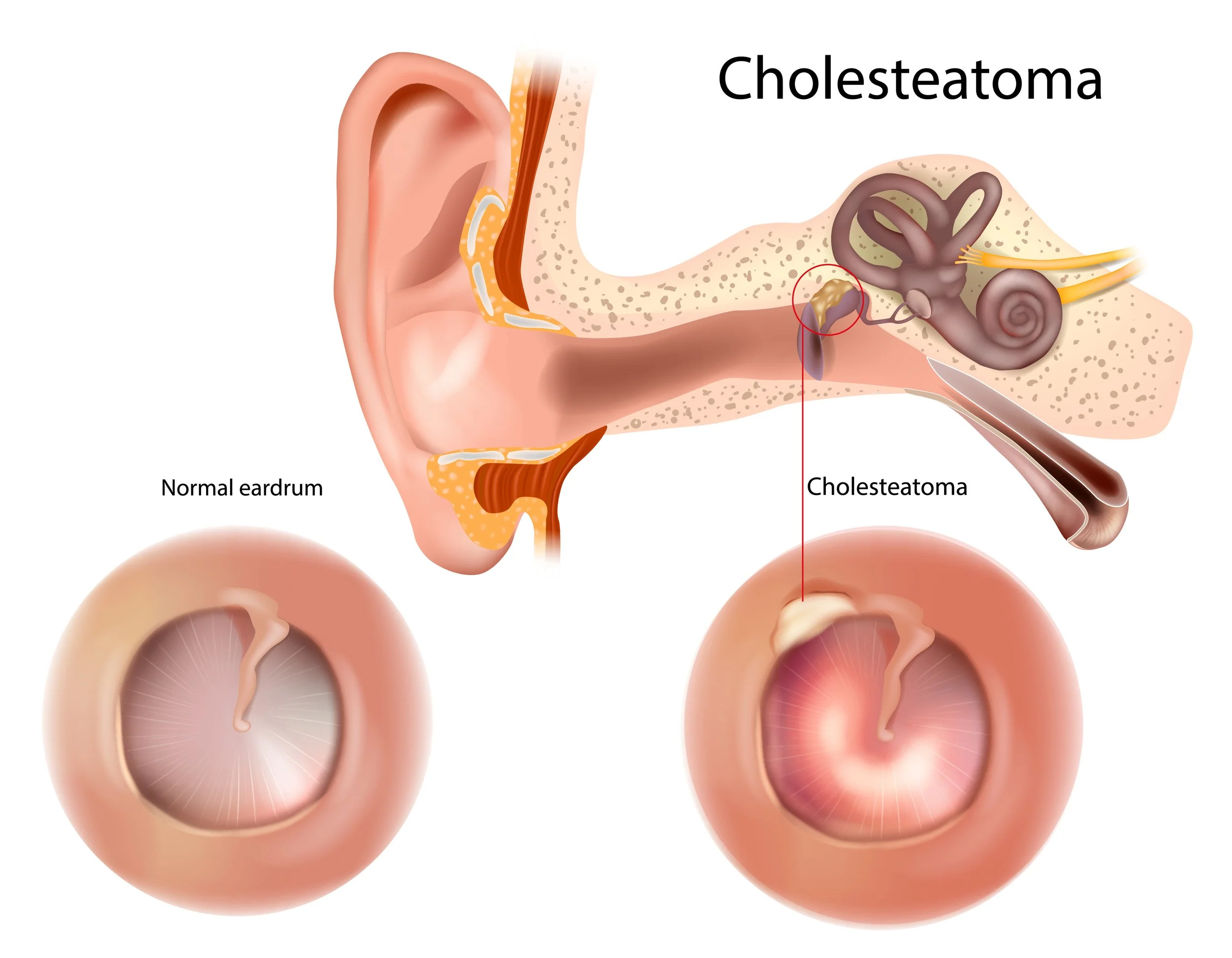
One complication of OM is a cholesteatoma, an abnormal accumulation of skin cells behind the tympanic membrane. This may result from AOM, chronic otitis media (COM), or tympanic membrane perforation. The treatment requires specialized ENT assessment and surgical intervention.
Extracranial Complications
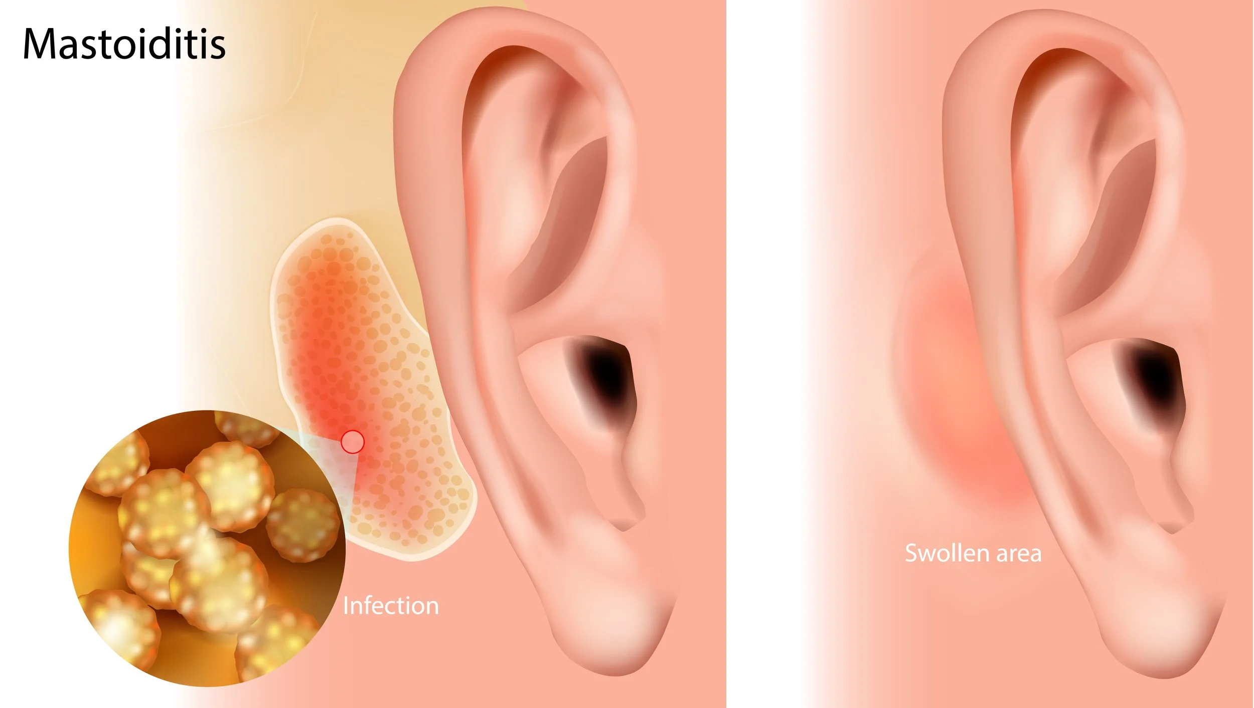
Extracranial complications involve extension of middle-ear disease outside the intracranial cavity. These infections may involve the mastoid air cells, causing acute mastoiditis; abscesses of the mastoid bone and periosteum (the bone's outer membrane); labyrinthitis (inflammation of the inner ear); and facial nerve inflammation, which may cause facial paralysis.
Intracranial Complications
More rarely, otitis media can be associated with intracranial complications. These serious and life-threatening infections include meningitis, brain abscesses, and venous sinus thrombosis. These infections require hospitalization, medical management by a multidisciplinary team, and possible surgical intervention.
Chronic Otitis Media Represents a Different Clinical Context
Acute and chronic middle-ear disease are not interchangeable when discussing complications.
Hearing Consequences Also Matter
Not every consequence of middle-ear disease extends an acute infection.
Middle-ear effusion may persist for weeks or months after AOM signs and symptoms resolve. Persistent middle-ear fluid reduces tympanic membrane mobility and can interfere with sound conduction. In these cases, hearing loss, speech delays, and developmental delays may occur. A hearing evaluation is warranted, with consideration for ENT referral.
Recognizing When the Clinical Picture Has Changed
Most uncomplicated AOM episodes do not result in serious complications. However, worsening symptoms or failure to improve as expected may warrant reassessment.
For appropriately selected children with non-severe AOM managed with initial observation, the American Academy of Pediatrics (AAP) guidelines recommend a plan to ensure follow-up and start antibiotic therapy if symptoms worsen or fail to improve within 48 to 72 hours.
The AAP guideline also recommends reassessing a child whose symptoms worsen or fail to respond to initial antibiotic treatment within 48 to 72 hours to determine whether therapy needs to be changed.
Consider persistent middle-ear effusion (OME) separately from persistent acute infection. OME is fluid in the middle ear without signs or symptoms of acute ear infection, and it may remain for weeks or months after the signs and symptoms of AOM resolve.
Why Accurate Assessment Matters
A child with middle-ear disease may have AOM, OME without signs or symptoms of acute infection, persistent middle-ear effusion following AOM, recurrent AOM, or chronic middle-ear disease. These conditions should not automatically be treated as equivalent.
Current clinical guidelines emphasize distinguishing AOM from OME. The AAP guideline states that clinicians should not diagnose AOM in children who do not have middle-ear effusion. Restated, a red tympanic membrane and no fluid identified on exam are not AOM.
The Bigger Picture
Serious complications should be kept in perspective. They are not the expected outcome of routine, uncomplicated acute otitis media. At the same time, studies of chronic disease and referral-center populations demonstrate that important extracranial and intracranial complications continue to occur.
For clinicians and families alike, the important message is balanced:
Most episodes of otitis media resolve without serious complications. However, persistent middle-ear effusion may affect hearing, and uncommonly OM may lead to extracranial or intracranial complications of middle-ear disease.
Distinguishing AOM from OME and recognizing persistent or atypical disease can support guideline-informed decisions about observation, treatment, hearing evaluation, and follow-up.
Medical Disclaimer
This article is provided for educational purposes only and is not intended to replace professional medical advice, diagnosis, or treatment. Patients and caregivers should consult a qualified healthcare professional regarding symptoms or treatment decisions.
Frequently Asked Questions
-
Most episodes of acute otitis media resolve without serious complications. Uncommon complications can occur when infection extends beyond the middle ear and may include mastoiditis, labyrinthitis, facial nerve involvement, meningitis, brain abscess, venous sinus thrombosis, and other extracranial or intracranial complications.
-
Yes. Middle-ear effusion can persist for weeks or months after the signs and symptoms of acute otitis media have resolved. Persistent fluid can reduce tympanic membrane mobility and interfere with sound conduction.
-
Middle-ear effusion associated with or following otitis media can interfere with sound conduction and contribute to hearing loss. When effusion persists, hearing evaluation and consideration of ENT referral may be appropriate.
-
Acute otitis media (AOM) and otitis media with effusion (OME) are not the same condition. OME involves fluid in the middle ear without signs or symptoms of an acute ear infection. Clinical guidelines emphasize distinguishing AOM from OME because their diagnosis and management differ.
-
For appropriately selected children with non-severe AOM managed initially with observation, AAP guidance recommends follow-up and starting antibiotic therapy if symptoms worsen or do not improve within 48 to 72 hours. Children whose symptoms worsen or fail to respond to initial antibiotic treatment within the same timeframe should also be reassessed to determine whether a change in therapy is needed.
References
1. Osma U, Cureoglu S, Hosoglu S. The complications of chronic otitis media: report of 93 cases. The Journal of Laryngology & Otology. 2000;114(2):97–100.
2. Penido NO, Chandrasekhar SS, Borin A, Maranhão ASA, Gurgel Testa JR. Complications of otitis media: a potentially lethal problem still present. Brazilian Journal of Otorhinolaryngology. 2016;82(3):253–262.
3. Ren Y, Sethi RKV, Stankovic KM. Acute Otitis Media and Associated Complications in United States Emergency Departments. Otology & Neurotology. 2018;39(8):1005–1011.
4. Nissen AJ, Bui H. Complications of Chronic Otitis Media. Ear, Nose & Throat Journal. 1996;75(5):284–292. PubMed PMID: 8935527.
5. Dongol K, Rayamajhi P, Gurung U. Complications of Acute and Chronic Otitis Media in a Tertiary Referral Center in Nepal. Turkish Archives of Otorhinolaryngology. 2020;58(4):234–240.
6. Rosenfeld RM, Shin JJ, Schwartz SR, et al. Clinical Practice Guideline: Otitis Media with Effusion (Update). Otolaryngology–Head and Neck Surgery. 2016;154(1 Suppl):S1–S41.
7. Lieberthal AS, Carroll AE, Chonmaitree T, et al. The Diagnosis and Management of Acute Otitis Media. Pediatrics. 2013;131(3):e964–e999. doi:10.1542/peds.2012-3488.